Deep Learning Models Can Detect COVID-19 in Chest CT Scans
The deep learning tools could distinguish between COVID-19 and other pneumonias in chest CT scans, enhancing coronavirus diagnosis.
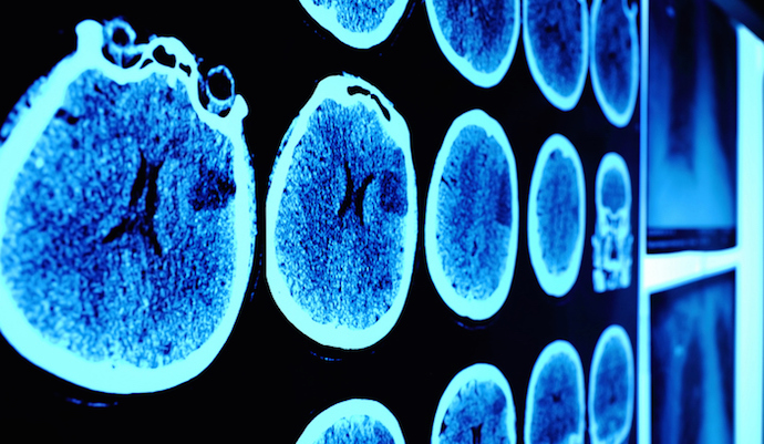
Source: Thinkstock
![]() By Jessica Kent
By Jessica Kent
- Deep learning tools were able to identify COVID-19 in chest CT scans, indicating that artificial intelligence could enhance diagnosis of the virus, according to a study published in Nature Communications.
For more coronavirus updates, visit our resource page, updated twice daily by Xtelligent Healthcare Media.
While CT scans have been useful in helping providers detect COVID-19, clinicians are discouraged from using these medical images for coronavirus diagnosis.
“CT evaluation has been an integral part of the initial evaluation of patients with suspected or confirmed COVID-19 in multiple centers in Wuhan China and northern Italy,” researchers noted.
“However, these guidelines also recommend against using chest CT in screening or diagnostic settings in part due to similar radiographic presentation with other influenza-associated pneumonias. Techniques for distinguishing between these entities may strengthen support toward use of CT in diagnostic settings.”
Because of the rapid increase in COVID-19 cases, artificial intelligence could play a role in detecting and characterizing COVID-19 on medical images.
“CT provides a clear and expeditious window into this process, and deep learning of large multinational CT data could provide automated and reproducible biomarkers for classification and quantification of COVID-19 disease,” researchers said.
Investigators from NIH and NVIDIA set out to develop and evaluate a deep learning algorithm to detect COVID-19 on chest CT using data from a globally diverse, multi-institutional dataset. The team obtained COVID-19 CT scans from four hospitals across China, Italy, and Japan, where there was a wide variety in clinical timing and practice for CT use in outbreak settings.
In total, researchers used 2,724 scans from 2,619 patients in this study. The study included two models that researchers used in series to come up with the COVID-19 final classification model.
The first model was a segmentation model that was used to define the lung regions which were subsequently used by the classification model. Initially, the team developed two classification models – one utilizing the entire lung region with fixed input size (full 3D), and one utilizing average score of multiple regions within each lung at fixed image resolution (hybrid 3D).
When distinguishing between COVID-19 and other conditions, the hybrid 3D model achieved validation accuracy of 92.4 percent, while the full 3D model achieved an accuracy of 91.7 percent.
To evaluate the utility of these models for COVID-19 sensitivity at independent institutions, researchers removed the cohort of COVID-19 patients from Tokyo, Japan from the training and validation datasets and re-trained the models using identical algorithm configuration and hyperparameters as the original models. Overall, validation and testing accuracy were stable between models trained with and without patients from the leave-out institution.
Because the models were able to distinguish between COVID-19 and other types of pneumonia demonstrates that there may be a role for AI as one element of a CT-enhanced diagnosis, researchers said. Subsequent models could include resource allocation, point of care detection for isolation of asymptomatic patients, or monitoring for response in clinical trials for medical countermeasures.
“Given the challenges in confidently distinguishing between COVID-19 associated pneumonia and other types of pneumonia, there may be a role for AI in CT-based diagnosis, characterization, or quantification of response,” researchers stated.
“Further work regarding the diagnostic utility of this algorithm in the setting of early vs. advanced COVID-19 related pneumonia is warranted.”
The researchers expect that this deep learning tool could be used in cases beyond the COVID-19 pandemic.
“While CT imaging may not necessarily be actively used in the diagnosis and screening for COVID-19, this deep learning-based AI approach may serve as a standardized and objective tool to assist the assessment of imaging findings of COVID-19 and may potentially be useful as a research tool, clinical trial response metric, or perhaps as a complementary test tool in very specific limited populations or for recurrent outbreaks settings,” researchers concluded.
Researchers have increasingly examined medical imaging and imaging analytics as a way to better diagnose COVID-19. The New England Complex Systems Institutes recently announced a partnership with the XPRIZE Pandemic Alliance to launch the COVID-19 CT Scan Collaborative.
The initiative aims to significantly accelerate the use of CT scans for COVID-19 diagnosis and treatment.
“CT scans can be a real game-changer in our global battle to end coronavirus,” said Yaneer Bar-Yam, PhD, President and Founder of the New England Complex Systems Institute, an independent academic research and educational institution.
“We need aggressive and bold actions to reduce transmission of COVID-19 to get ahead of the outbreak so that it is stopped. It will take the global community to accelerate how we meet these challenges.”